CellChek® XL & SL
Gently Used Gold Standard Specular Microscopes (Limited Availability)
Endothelial cell morphology is like a ‘canary in the coal mine’; a potential warning that the structural stability of the endothelium has been affected, possibly by surgical procedures, disease, trauma or contact lenses.

Clinical Applications
Corneal disease management.
Contact/specialty contact lens fittings.
Routine eye care
Clinical Benefits
Identify pre-existing low density and dystrophies that may affect positive surgical outcomes
Confirm recommended cell density/morphology for scleral/specialty contact lenses
Regulatory
FDA 510(k) Cleared | CPT Code 92286
FDA 510(k) Cleared Database
Health Canada Licensed
Gently-used devices available in the USA only
CellChek® SL | XL Models
Gently used devices come with ‘same-as-new’ warranty. Examples include trade show floor / demo units, clinical trial returns and trade-ins. Limited availability. Only available in the USA.
***Please note that Konan does not buy and sell used equipment.***

Clinical Benefits
Cataract Surgery and
Premium IOLs
Low endothelial cell counts and pre-existing dystrophies can markedly reduce the potential for positive surgical outcomes from an otherwise uneventful cataract surgery. Surgeons are finding these data points critical when recommending premium IOLs: verify and document pre-operatively that the cornea is not suspect to more likely post-op complications difficult to explain with the investment in premium IOLs.
General Corneal Health Assessment
Konan specular microscopy is an invaluable tool to screen for corneal diseases as such as Fuchs’ Dystrophy, keratoconus, other corneal dystrophies, and trauma. You won’t believe what you’ve been missing.
Refractive Surgery
Contact Lenses
It is not a coincidence…

Robust Analysis Methods
It all starts with robust data assessment of even the most difficult cases. Fully automated analysis is quick and easy, but Konan exclusively has options to maximize the sparse information on even advanced disease-state corneas.
FDA Cleared Database
Konan CellChek® systems include robust database functions. Corneas with advanced disease states may be reanalyzed using more than one analysis strategy when there are few cells visualized and may be reassessed at a later date without overwriting original data.
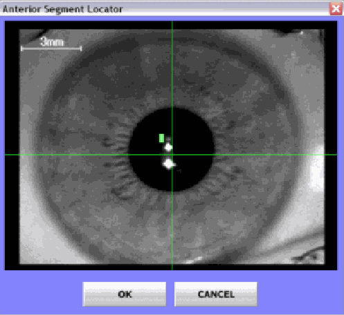
Location, Location, Location
Easy location identification. Fixation direction is not enough – you cannot draw any conclusions about change over time unless you know the sample location.
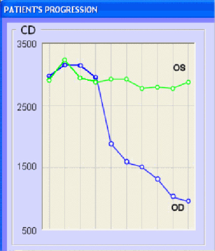
Trends Analysis
Clear assessment of changes over time. Statistically valid trends can only be obtained if you are comparing same data locations.
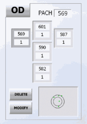
Multi-Point Pachymetry
FDA Cleared Hardware & Software
Easy location identification. Fixation direction is not enough – you cannot draw any conclusions about change over time unless you know the sample location.
DICOM
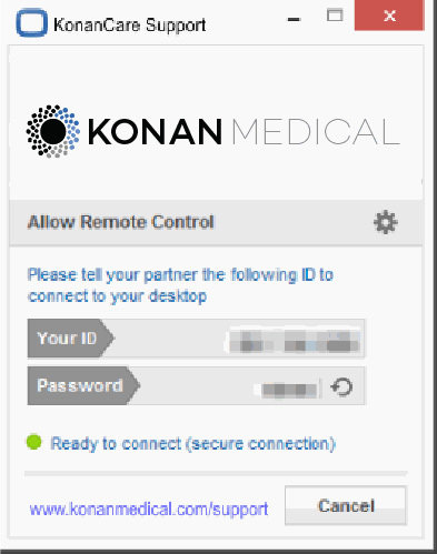
KonanCare
Scientific and Clinical Publications

As a corneal specialist for the last 22 years I would feel incomplete without my ability to perform specular in the clinical and eye bank setting. I have Konan units from the earliest to the most recent, all in service currently. They are reliable, accurate, and serve an essential role in my clinical practice and eye bank operations. The service delivered by Konan is some of the best I have experienced in the ophthalmic field. Specular microscopy provides a superb method of clinical screening for disease, an educational training tool for patients, their families and office and eye bank personnel, as well as a tissue evaluative method for eye banking. It is time and cost effective. I wouldn’t want to practice without a Konan specular microscope.
George O. Rosenwasser, MD

We utilize the Konan specular microscope after all of our DSAEK surgery. The information that the specular microscope provides is valuable for assessing the long term effects of the surgical trauma of the procedure, and specular microscopy is critical to understanding your personal outcomes with DSAEK.
Mark Terry, MD

I have been surprised at how many of my cataract patients had substantial endothelial dystrophic changes that are not readily visualized through the slit lamp but detectable with specular bio-microscopy. Konan endothelial analysis is now an important part of the work up for all of my patients needing cataract surgery, especially for my premium IOL patients. It is important for us to educate our patients about their pre-existing endothelial conditions so they are fully prepared for the surgery and sometimes the prolonged postoperative corneal edema and blurriness as a result of corneal endothelial sub-function. As one doctor once said: “If you mentioned a complication before the surgery and it did occur, then you are a great doctor as having predicted it; but, if you failed to mention a complication before the surgery and it did occur, then you are a bad doctor in having caused it. Not only for doing a better job in the management of patient expectations post operatively, but also and most importantly providing a top qualify and comprehensive eye care for the cataract surgery patients including with premium IOLs with their rising expectations today, specular microscopy is now in my opinion a requisite contemporary tool for all cataract surgeons who want to provide the state-of-the-art refractive cataract surgery for their patients.
Ming Wang, MD, PhD

From the outside it would appear that I have a ‘healthy’ patient base of young and middle aged adults. In reality, I have an entire generation of adults who have been wearing low Dk hydrogel lenses for two-thirds of their lives. Every day I see the results of poor lens breathability combined with even worse lens hygiene and discard habits. My specular microscope gives me the information I need to educate patients on why we need to alter their lens care regimen or change their lens brand and material, even though they may be asymptomatic and ‘perfectly happy’ in their prior lenses.
Stacie Virden, OD

My Konan specular microscope is the best investment that I have ever made.
Steven Bovio, OD

I never realized what I was missing in my corneal examinations until I was able to view images with my Konan specular microscope. Our practice has implemented this phenomenal technology in all of our contact lens exams. We can monitor endothelial changes not only in extended wear patients but in traditional hydrogel wearers who may need to be in SiHys. The Konan specular microscope can detect early endothelial damage and allow us to modify patient behavior or lens fit as necessary. We can now more accurately monitor our Fuchs patients and patients with early guttata or endothelial changes. Scanning a patient’s endothelium has also now become standard pre and post cataract surgery especially if we are considering implanting a multifocal IOL. We can detect any early endothelial damage which may limit optimal post-operative VA. It also allows us to monitor any endothelial damage post-operatively as there is a small amount of endothelial cell loss post cataract surgery. The favorable reimbursement has allowed us to be profitable while still being able to offer our patients the latest technology available.
Thomas P. Kislan, OD

A comprehensive anterior segment exam, must include corneal endothelial evaluation. The Konan system takes specular microscopy to the next level, and allows for a swift and accurate appraisal of the most challenging corneas. In a busy clinic, ancillary testing that can easily be performed by support staff is invaluable, and all of my technicians are well versed in the use of the Konan specular microscope. This is a no brainer for cornea specialist, but the cataract surgeon performing lifestyle lens implantation should know the health of the corneal endothelium when recommending a lens like a multifocal. Furthermore, the addition of pachymetry provides a bridge between anatomy and physiology of the endothelium and secondary detergesence.
Jonathan D. Solomon, MD

Konan specular microscopes are recognized in the industry as the de facto standard for ECD determination. This is supported in the minutes from the FDA panel meeting to discuss the ARTISAN® lens: “Data from 12 sites were chosen because they used the Konan specular microscopes. This instrument is now the accepted standard for the most accurate determination of endothelial cell density.
R. Doyle Stulting, MD, PhD.

The Konan Specular Microscope is very tech friendly. It is easy to use and the information is outstanding. Managing corneal dystrophies and predicting outcomes, such as when to perform a DSEK procedure, has been very advantageous in the care of my patients. The ability to explain subtle visual acuity decrease has helped reduce patient anxiety. The Specular Microscope is well worth the money.
John Coble, OD, FAAO

My offices have had Konan endothelial microscopes for about 3 years. The Konan specular microscope allows me to quantify my patients corneal health in a very easy to understand manner. Not only does Konan specular microscopy give great clinical information it also paid for itself well within the first year. For Doctors who see many contact lens patients the specular is a great adjunct to the Topographer and in many ways is a much better predictor of contact lens success. I find the Konan specular microscope is most helpful for predicting future success with the overwear contact lens patient.
Kerry Gelb, OD

Vague vision complaints from the elderly and long term contact lens patients are commonly heard in a busy practice. Morning blurred vision, nighttime glare, or reduced contact lens wear time are complaints that have been heard over and over. Sometimes, patients assume that their problems are too small to discuss, and unless we are able to phrase our questioning appropriately, or we are able to observe minute endothelial changes under a slit lamp, the cause and resolution can remain elusive. Until now! The Konan specular microscope (KSM) has been able to help provide part of the solution. Endothelial changes that were not detected even with the best slit lamp images can now be documented with the KSM. Once detected, these symptoms and complaints can often be reduced or eliminated. Whether it means the use of a hyperosmotic drop or a contact lens refit to a higher oxygen transmitting lens, patients complaints decrease as they are able to maintain clearer vision or achieve longer, more comfortable contact lens wear. The KSM has become an important part of my total patient care.
Jeffrey E. Schultz, O.D., M.S., F.A.A.O.

Simply an indispensable technology for Ophthalmology and Optometry, from Konan Medical, the “Apple” of ophthalmic device companies.
Paul Karpecki, OD, FAAO

We bought a new N**** in 2017 but it was very difficult to use, temperamental, and had poor image capture and analysis. After months of fruitless attempts to improve things, we decided to switch to Konan CellChek and have been much happier with its capabilities and ease of use.
Bala Ambati, MD
Clinical Resources
If a Patient Complains of Contact Lens Intolerance, Don’t Forget to Check the Endothelium
Karpecki, P. Practice Pearl of the Week. Review of Optometry (2011).
Endothelial Cell Density to Predict Endothelial Graft Failure After Penetrating Keratoplasty.
Lass, J. H., Sugar, A., Benetz, B. A., Beck, R. W., Dontchev, M., Gal, R. L., … & Stulting, R. D. (2010). Endothelial cell density to predict endothelial graft failure after penetrating keratoplasty. Archives of ophthalmology, 128(1), 63-69.
Continuous evolution of endothelial keratoplasty changes field of corneal transplantation
John, T. Continuous evolution of endothelial keratoplasty changes field of corneal transplantation. Ocular Surgery News (2013).
Use Specular Microscopy to Diagnose Corneal Disease
Craig, T. “Use specular microscopy to diagnose corneal disease.” Review of Optometry (2009).
Fundamentals
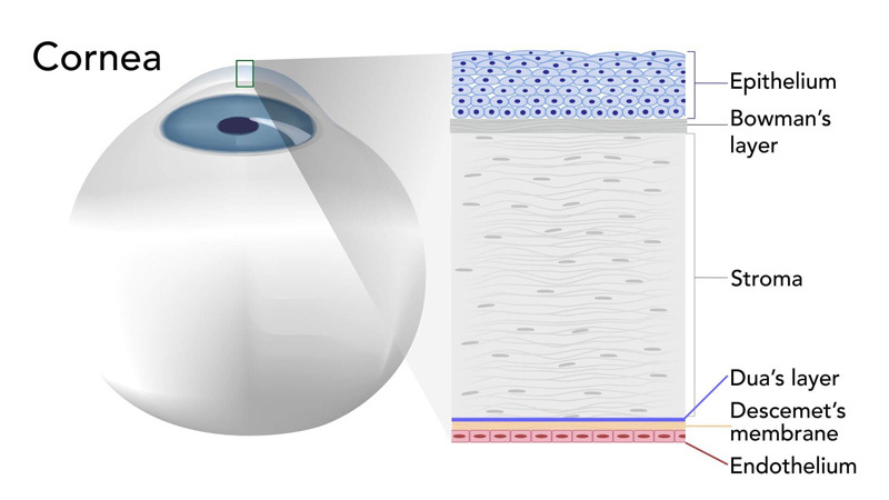
The Cornea
The corneal endothelium is a single layer of cells whose function is to maintain the balance of fluid (aqueous) within the cornea by means of a barrier effect, and to remove excess aqueous from the cornea by means of a pumping mechanism. A properly functioning endothelium maintains the correct clarity and shape of the corneal required for clear vision. When endothelial cells are lost or damaged, the remaining cells grow in size and change shape to fill in the gaps in order to maintain structural integrity. If too many cells are lost or damaged, the pumping mechanism may be negatively affected, resulting in corneal edema which may lead to partial or complete loss of vision.
Cell Density & Morphology Changes
Endothelial cells may be lost due to traumatic injury, damage during eye surgery (corneal incisions, phaco-energy), laser surgery, intraocular lenses, pharmaceutical agents contained in eye drops, or simply through the aging process. The number of cells along with the variance of the sizes and shapes of the endothelial cells serve as quantitative and qualitative indicators of the health of the cornea. With the prevalence of corneal and anterior segment surgeries, implantable eye devices, and contact lenses, the value of monitoring the corneal endothelium has never been higher. Konan’s specular microscope makes it possible to observe the endothelium at high magnification and provides a detailed assessment of the cornea. This important information helps define the best mode of surgery or therapy for a given patient.
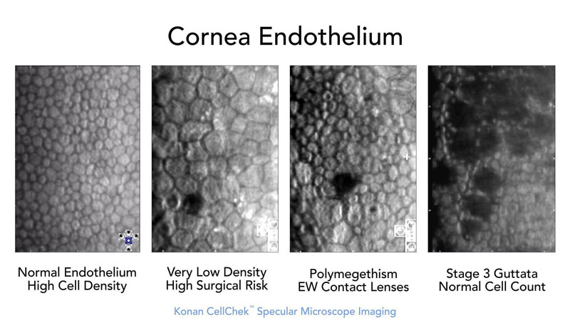
Endothelial Morphology Examples
Normal Endothelial Cell Density (ECD)
Normal ECD declines with age.
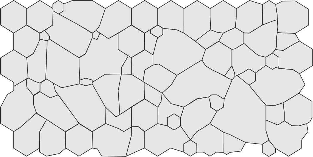
Low ECD
Low ECD is a risk factor for cataract and corneal surgeries that can be missed without the use of specular microscopy.
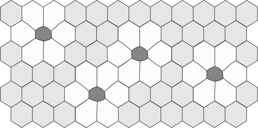
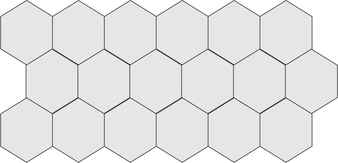
Polymegethism
Change from uniform cell sizes to variable cell sizes is an indication of distress to the endothelium.
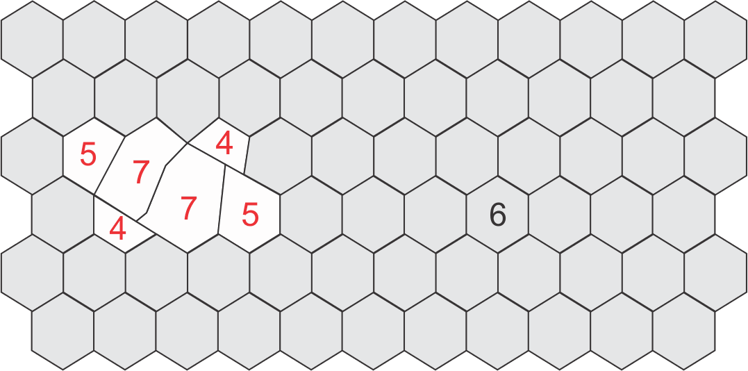
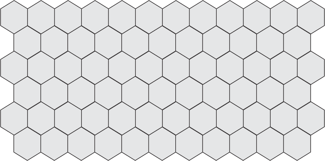
Pleomorphism
Change from optimum hexagonal cell geometry to variable / compromised cell shapes.
Normal ECD with Polymegethism and Pleomorphism
Here the ECD is typical, but there are also large changes in morphology with high variance in cell area / sizes (pleomorphism) and a large number of non-hexagonal cell shapes (4, 5, 7 sided cells, i.e. pleomorphism).
Konan Medical is trusted to provide the most widely used specular microscopy – endothelial analysis tools for routine clinical practice, clinical trials, professional reading centers, and the world’s #1 eye bank system for qualification of donor corneal tissue.
This is not a coincidence, it has been and is continuously earned… only from Konan.
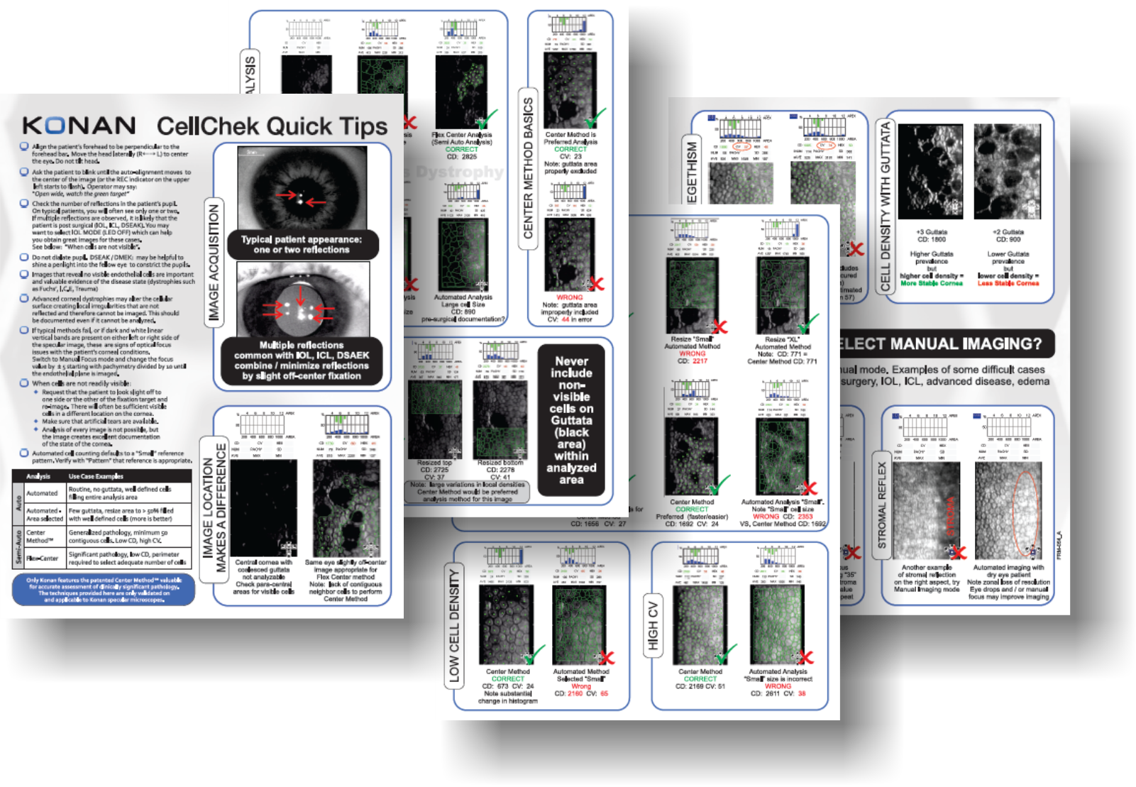
Konan CellChek Quick Tips
Konan CellChek system has long been the market leader for robust endothelial analysis. But it is the difficult cases where we really shine. Contact Konan to obtain a copy of CellChek “Quick Tips” for in-depth clinical pearls for assessment of your most challenging cases including those that are good images of bad corneas.
ICL | PIOL | Scleral Lenses

Vance Thompson, MD
“I use the Konan specular microscope to measure every phakic lens candidate. It helps me to ensure they have an adequate endothelial cell count for their age.”

Theodore Perl, MD
It is important to select patients whose endothelial cell density and morphology are normal.
Avoiding Complications with Phakic IOLs” (Cataract and Refractive Surgery Today, October 2009)
ICL (implantable contact lens) or PIOL (phakic intraocular lens)
An endothelial cell count to verify that a patient’s cornea has an adequate endothelial cell density is an FDA labeling requirement for patient screening and selection prior to implantation of phakic intraocular lenses (Intraocular Contact Lenses, or “ICLs”). Without an adequate endothelial cell count, the current ICLs are contraindicated for implantation and clearly highlights the importance of this screening procedure. Endothelial cell density must be accurately checked preoperatively to ensure that a patient is a suitable candidate, and then should be monitored periodically postoperatively to verify the endothelium’s continued health. Although an endothelial cell count can be crudely estimated using a card developed in the 1980′s, this technique has significant limitations that are now well answered with Konan’s specular microscopy technologies:
- High precision and repeatability
- Excellent visualization of even stage +1 / +2 guttata that could significantly skew results
- Analysis of both cell counts and cell morphology changes (morphology is not assessed with a “card”)
- Changes in morphology can be a very sensitive indicator of corneal stress
- Unique ability to identify endothelial data location and assess trends over time
- Acquiring endothelial cell count or morphology changes over time from dissimilar locations can present erroneous assumptions on trends analysis
- Technician administered diagnostic test with full photographic documentation and statistics in only seconds.
- Does not require additional physician chair time and eliminates the inherent problems of “card” estimates.
- Important complementary uses for pre-operative assessments of corneal and other anterior segment procedures
ICL Products
Premium refractive products require premium attention to eligibility. Konan CellChek® provides easy to use yet robust analytics to assure proper patient selection. There is a reason major manufacturers of ICL’s use Konan specular microscopes for collection of FDA safety data. Your patients deserve no less.

Visian ICL™ - Staar Surgical™
FDA Professional labeling including ECD minimum requirements:

Verisyse™ - Abbott Medical Optics™
FDA Professional labeling including ECD minimum requirements:

AcrySof® Cachet® - Alcon®
Scleral Lenses with High Risk Endothelial Cell Density
Fundamentals of the importance of endothelial cell density to support implantation of phakic IOLs described above, may have parallels in decision making for scleral lens therapies.

Christine Sindt, OD, FAAO
Clinical Professor Ophtalmology and Vision Sciences, University of Iowa
It is common to experience corneal edema with endothelial cell counts under 800 cells/mm².
Navigating the Slippery Slope (Review of Optometry, April 2015)

Eef van der Worp, BOptom, PhD, FAAO, FIACLE, FBCLE, FSLS
Pacific University, Oregon | University of Maastricht, Netherlands
Endothelial cell count of less than 800 cells/mm² is where the problems may arise (Sindt 2010a), and endothelial cell counts <1,000 cells/mm² should be handled with extra care and should not be fitted with scleral lenses to avoid edema.
A Guide to Scleral Lens Fitting

Product Specifications
| Model no. / Product name | CellChek® 20-1 or CellChek® 20-1 PLUS / Konan Specular Microscope XX |
| Type | Class I, Type B |
| Photographic field | 0.25 x 0.55mm |
| Photographic capability | Automatic |
| Photographic location | Center, Peripheral locations (0, 45, 135, 180, 225, 315 degrees) |
| Central corneal thickness | Measurable |
| Illumination | LED |
| Analyzing method | Auto Center Method / Auto F Center Method / Auto Trace Method / Center Method / F Center Method / Trace Method |
| Analysis data | Cell density (mm2), Coefficient of Variation, Standard Deviation, Percentage of hexagonal cells, Numbers of calculated cells, Average cell area (µm2), Maximum cell area (µm2), Minimum cell area (µm2), Distribution of number of sides histogram (%), Distribution of area histogram (%) |
| Integrated monitor | 10.1” Capacitive touch panel with a 180 degrees flexible horizontal & vertical movement capability. |
| External interface | EMR connection / DICOM capability |
| External ports | USB3.0 x 4 Type A , RJ-45 |
| Power consumption | 100 VA |
| Dimensions | 310 (W) X 459 (D) X 451 (H) mm (with the monitor at the rear-side) |
| Weight | 19.6 kg |
What Konan Medical Customers Say

We utilize the Konan specular microscope after all of our DSAEK surgery. The information that the specular microscope provides is valuable for assessing the long term effects of the surgical trauma of the procedure, and specular microscopy is critical to understanding your personal outcomes with DSAEK.
Mark Terry, MD
Devers Eye Institute, Portland, OR USA

The Konan Specular Microscope is very tech friendly. It is easy to use and the information is outstanding. Managing corneal dystrophies and predicting outcomes, such as when to perform a DSEK procedure, has been very advantageous in the care of my patients. The ability to explain subtle visual acuity decrease has helped reduce patient anxiety. The Specular Microscope is well worth the money.
John Coble, OD, FAAO
Eyecare of Greenville, Greenville, TX USA

I never realized what I was missing in my corneal examinations until I was able to view images with my Konan specular microscope. Our practice has implemented this phenomenal technology in all of our contact lens exams. The favorable reimbursement has allowed us to be profitable while still being able to offer our patients the latest technology available.
Thomas P. Kislan, OD
Medical Director, Hazelton and Stroudsburg Eye Specialists, Hazelton, PA USA

My offices have had Konan endothelial microscopes for about 3 years. Not only does Konan specular microscopy give great clinical information it also paid for itself well within the first year.
Kerry Gelb, OD
Contact Lens and Vision