
objectiveFIELD®
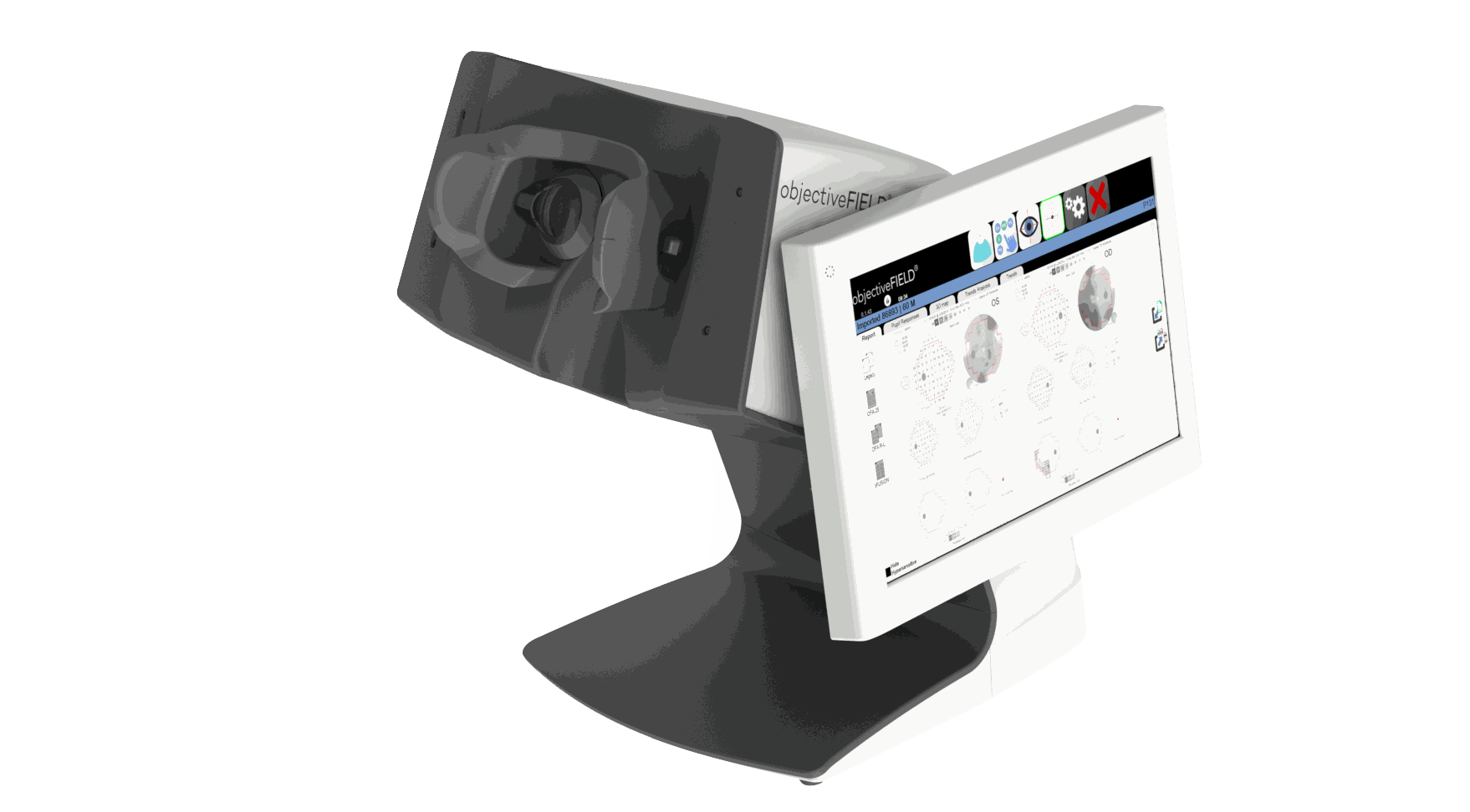
objectiveFIELD® is the first and only truly objective perimeter that provides sensitivity and latency data.
Like multi-focal VEP, but no electrodes!
Overview
 The objectiveFIELD Analyzer (OFA®) utilizes patented mfPOP technology, which is like multi-focal VEP (mfVEP), but without the need for electrodes.
mfVEPs (and mfERGs) and OFA measure response size over time. Thus, all three give a peak response (sensitivity) and a time-to-peak (latency or delay) that are the averages of many presentations at each tested location. When tested head-to-head OFA and mfVEPs agree.1
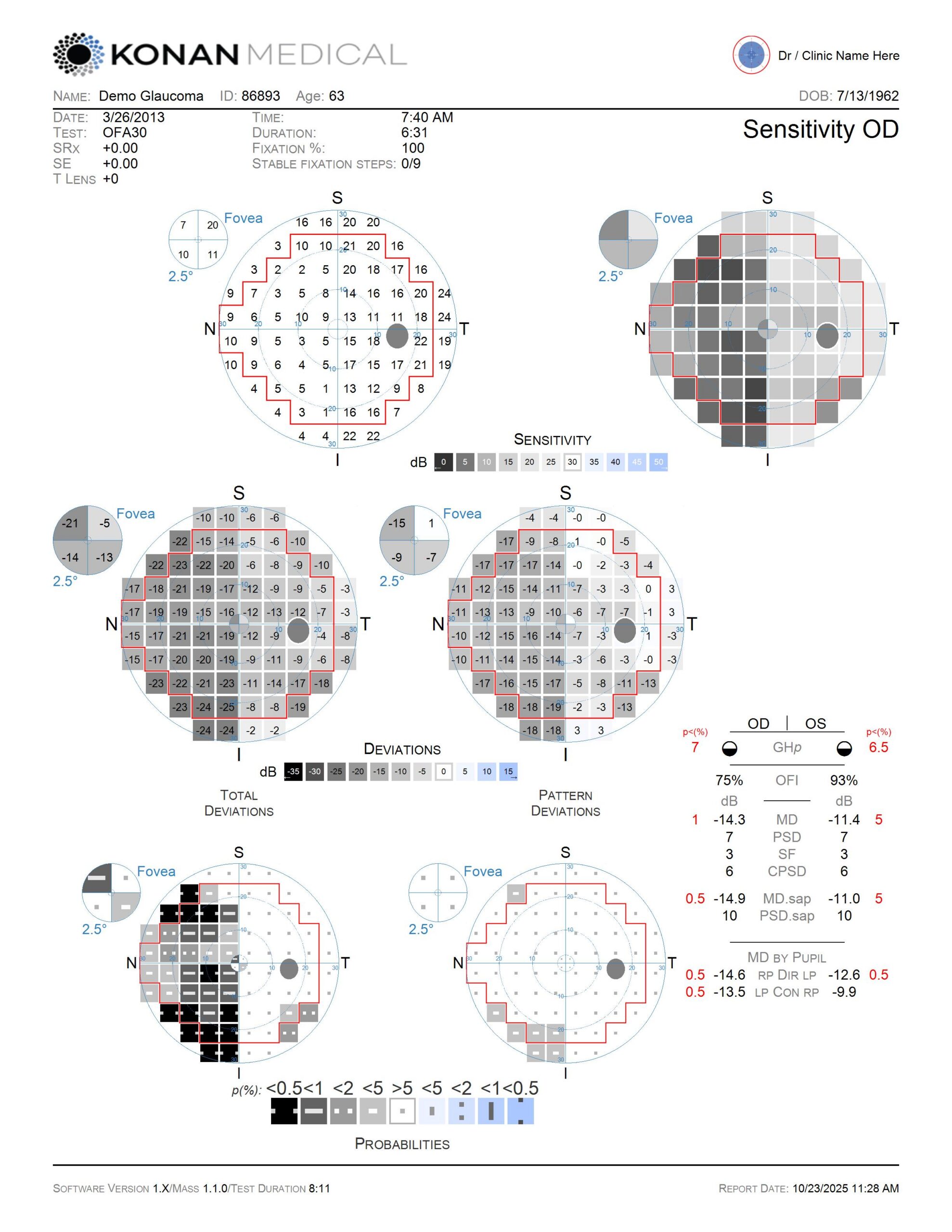
Similar to mfVEP data, but with familiar, standardized perimetry-style reports, OFA maps spatially-resolved amplitude data (Sensitivity in decibels) and latency data (Delay in milliseconds), producing four visual field reports per exam, in about 4 minutes per eye.
1. Sabeti F, James AC, Carle CF, Essex RW, Bell A & Maddess T. (2017). Comparing multifocal pupillographic objective perimetry (mfPOP) and multifocal visual evoked potentials (mfVEP) in retinal diseases. Sci Reports 7(45847): 1-12; DOI: 10.1038/srep45847.
What is delay data (latency) and why should you care about it?
Delay is an additional measure of visual function that is independent of Sensitivity. While well understood in visual electrophysiology, this concept is of course, previously unheard of in perimetry.
Both sensitivity and delay give similar shaped visual field features (field defects) linked to nerve-fibre bundles in glaucoma, but they can occur in different parts of the field, and one can occur without the other, possibly reflecting different stages of the disease.
There is evidence that SAP sometimes incorrectly identifies a delay defect as a sensitivity defect. In that case the SAP field and the OFA delay field match, and both agree with OCT.2,3
Most importantly, because some eyes progress on delay alone, clinicians can identify more eyes that are progressing by monitoring delay fields.
2. Thepass G, Lemij HG, Vermeer KA, van der Steen J, et al. Slowed saccadic reaction times in seemingly normal parts of glaucomatous visual fields. Front Med (Lausanne) 2021;8:679297. 10.3389/fmed.2021.679297
3. Maddess T, Carle CF, Kolic M, Essex RW, et al. Diagnostic power and reproducibility of objective perimetry in glaucoma. J Glaucoma 2024;32:940-950. 10.1097/IJG.0000000000002485
Single-Axis vs. Dual Axis Testing: What is it and why should you care about it?
Click here to download OFA Fundamentals and learn more.
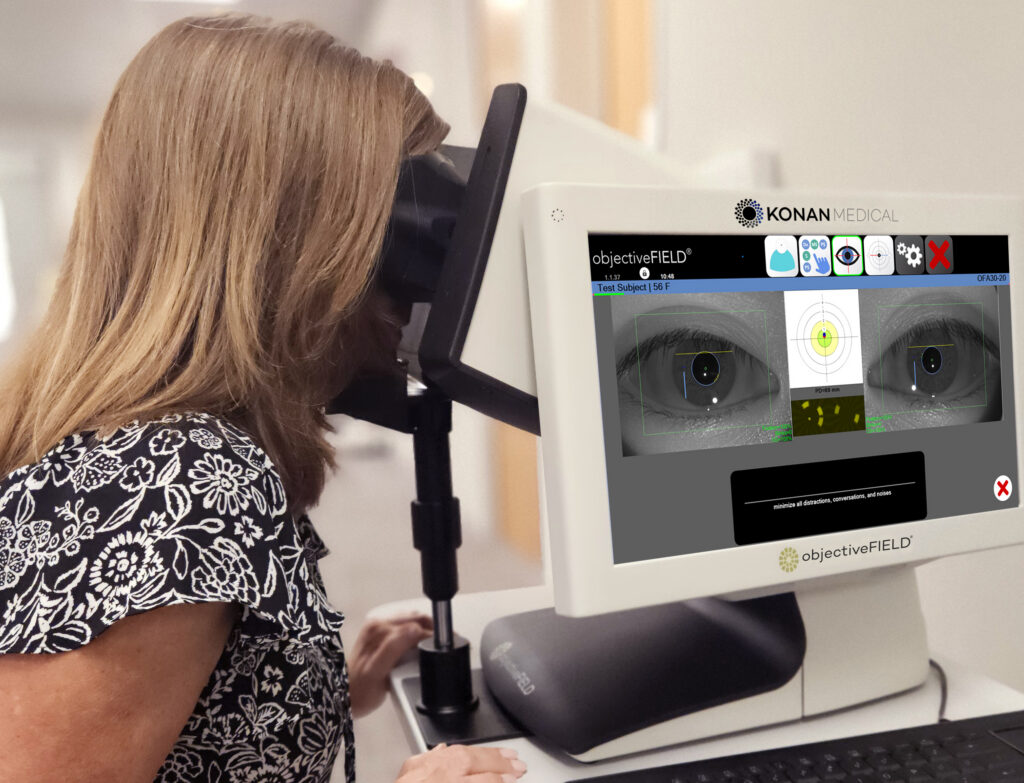
Easy on patients and staff: The objective, bilateral exam is easy to administer and well tolerated by patients. Normal blinking during the exam is allowed.
Both eyes are tested individually, but concurrently (a dichoptic presentation).
- Objective: No button to push, no cognitive / motor response required
- Bilateral: Both eyes tested at the same time, no eye patch
- Novel stimuli: Large, blurred, yellow stimuli (no blue), is less susceptible to refractive error and lens brunescence from cataracts
- Easy to administer: Monitor patient compliance in real-time

Clinical Applications
Diabetes, AMD, glaucoma
neuro-ophthalmology.
objectiveFIELD is not FDA cleared for the specific diagnosis of any condition.

Clinical Benefits
.Completely objective,
no button to press.
Sensitivity & delay data.
Bilateral exam, no eye patch needed.
Good correlation of glaucomatous RNFL loss with visual field loss

Regulatory
CE Marked
Health Canada Licensed
Pharmaceuticals and Medical Devices Agency (Japan)
What Visual Field Test Protocols Are Available?
What Does objectiveFIELD® Measure?
Novel stimuli are presented to each eye individually, interleaved, with non-overlapping timing.
Cortically mediated direct and consensual pupillary responses are recorded by video cameras under infra-red light using sophisticated pupillography (not gaze tracking / saccades / visual grasp etc.).
Sensitivity is measured objectively from the amplitude of the pupillary responses (relative change in pupil diameter) which is converted to the familiar decibel scale.
Delay (latency / time-to-peak constriction of the pupillary responses) is also measured objectively, in milliseconds, which is new information not available in SAP/VR style devices.
The result is four visual fields per exam.
- OD sensitivity
- OS sensitivity
- OD delay
- OS delay
Click to Download OFA Fundamentals
Standard Perimetry Reports
Ted Maddess, PhD, discusses the objectiveFIELD Analyzer at the American Academy of Ophthalmology
Scientific and Clinical Publications
How does objectiveFIELD work?
How long does an exam take?
High-Resolution test-protocols (30° & 15°) take approximately 8 1/2 mins, for both eyes, concurrently. These tests are administered as 9 shorts steps (~40 seconds each / 6 1/2 mins actual test time). The patient can take a short break between steps while the system checks the reliability of the data collected in each step. Cadence of the exam is controlled by the operator.
High-Efficiency test-protocols (30° & 10°) take only 90 seconds, also for both eyes, administered as one continuous 90 second step.
How does objectiveFIELD compare to standard automated perimetry?
objectiveFIELD® (OFA®) is 100% objective and utilizes a dichoptic presentation stimulus. It is easy on patients and technicians because there is no button to push and both eyes are tested at the same time.
Responses are measured objectively using a patented “Clustered Volley”, multifocal pupillographic objective perimetry method (mfPOP). OFA® stimuli covers more than 99% of the 30-2 retinal area, while SAP Goldman III stimuli only samples approximately 0.5% of the 30-2 area with the assumption that the remaining 99.5% of the non-sampled central area is well represented by the small stimuli.
Can the device accommodate smaller faces?
The human interface design is designed for a wide range of human morphology. Minimum IPD is ~ 42mm.
Is refractive correction required?
OFA® stimuli are much larger than Goldmann III stimuli and have blurry edges, and are therefore designed to be less susceptible to refractive errors than SAP. Stimuli are presented at optical infinity. Aspheric trial lenses are provided in 3 diopter steps for larger refractive errors. The system includes a calculator that specifies the correct trials lenses (spherical equivalent) by entering the patient's spectacle Rx.
Product Specifications
| Fundamental Method | Multifocal pupillographic objective perimetry (mfPOP) |
| Intended Clinical Purpose and Use | Visual field examination to measure visual field abnormalities |
| Visual System Stimulus | Patented clustered volley, sparse, multifocal stimulus |
| Visual Function Assessment | Regression-based multifocal analysis |
| Visual Field Defect Assessment | Population sample normal database comparison |
| Test Options | 30°, 24°, 15°, 10° tests |
| Trial Lenses | +3D, -3D, +6D, -6D, +9D, -9D |
| Background Brightness | 10 cd/m2 |
| Regulatory | FDA 510(k) Cleared K063310 | CE Marked | Health Canada Licensed
Pharmaceuticals and Medical Devices Agency (Japan)
|
| Instructions for Use (IFU) | Download |
Intended Purpose of Device & Indications for Use
This device is not intended to diagnose any disease. Use of the device in such a manner may result in misdiagnosis of serious pathologies. It is solely the physician’s or operator’s responsibility to interpret the test results.
The intended use of the device is to aid in the measurement of visual field abnormalities.
The device is intended for use by healthcare professionals or paraprofessionals in either a professional healthcare environment or other places where patients are present.
objectiveFIELD® should not be used to examine the following category of patients, this category may include, but not be limited to: patients with irregular shaped and stationary pupils; patients with Horner’s syndrome or other disorders affecting pupil response; patients with unusually small pupils or patients treated by drugs that would affect the pupil reactivity.
What Konan Medical Customers Say

The service delivered by Konan is some of the best I have experienced in the ophthalmic field.
George O. Rosenwasser, MD
Central Pennsylvania Eye Institute – Medical Director, Gift of Life Donor Program Eye Bank

Simply an indispensable technology for Ophthalmology and Optometry, from Konan Medical, the “Apple” of ophthalmic device companies.
Paul Karpecki, OD, FAAO
Clinical Director, Kentucky Eye Institute
Chief Clinical Editor, Review of Optometry
Associate Professor, Kentucky College of Optometry

Thank you to Konan for transforming eye disease detection and patient care!
Glen R. Owen, OD, FAAO
Edmonds Eyecare Associates, WA

My offices have had Konan endothelial microscopes for about 3 years. Not only does Konan specular microscopy give great clinical information it also paid for itself well within the first year.
Kerry Gelb, OD
Contact Lens and Vision